Plain-language answer
Diabetic macular edema, or DME, is swelling in the center of the retina caused by diabetes-related leakage from damaged blood vessels. It is one of the most common causes of blurred central vision in diabetic eye disease and is often treatable when diagnosed early.
What it is
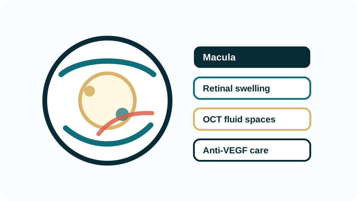
The macula is the part of the retina used for reading, driving, and detailed vision. In DME, fluid collects in or under the macula, thickening the retina and reducing the quality of central vision. Some patients notice blur or distortion, while others are diagnosed by OCT before symptoms become severe.
Symptoms
Symptoms can include blurry vision, wavy vision, reduced contrast, difficulty reading, washed-out colors, or vision that fluctuates during the day. DME may affect one eye more than the other, and the better-seeing eye can mask symptoms until disease is advanced.
Diagnosis
OCT imaging is central to DME diagnosis because it measures retinal thickness and shows fluid pockets. A dilated exam, fundus photography, and fluorescein angiography may also be used to assess leakage, blood flow, and the broader stage of diabetic retinopathy.
Treatment options
Anti-VEGF injections are commonly used first for center-involving DME. Some patients may need monthly injections initially, followed by a personalized interval based on OCT response, vision, and fluid activity. Steroid therapy or focal laser may be considered in selected cases, especially when inflammation, chronic leakage, or treatment response patterns suggest a different approach.
Follow-up and expectations
DME is usually managed over time rather than fixed with one visit. The goal is to reduce swelling, stabilize or improve vision when possible, and prevent worsening. Control of blood sugar, blood pressure, kidney disease, and cholesterol can also affect outcomes.
Why choose Retina of North Texas for DME
DME treatment is highly individualized because two patients with the same diagnosis can have different OCT patterns, vision levels, treatment responses, and systemic risk factors. Dr. Chavala evaluates the amount and location of fluid, the chronicity of swelling, prior treatment response, and coexisting diabetic retinopathy before recommending a plan.
The practice emphasizes clear communication about why injections may be needed, how progress is measured, and when treatment intervals can be adjusted. Same-clinic-day access is available when possible for sudden worsening, and follow-up is structured around OCT findings rather than a one-size-fits-all schedule.
Because DME can recur after it improves, patients are given a practical follow-up plan and clear instructions about symptoms that should prompt earlier evaluation. This helps avoid treatment gaps that can allow fluid to return and central vision to decline.
For patients who need injection treatment, Dr. Chavala has extensive high-volume experience, with >99% safety rate for eye injections in practice experience.
Frequently asked questions
Is diabetic macular edema the same as diabetic retinopathy?
DME is a specific complication of diabetic retinopathy that causes swelling in the macula. A patient can have diabetic retinopathy with or without macular edema.
How is DME treated?
Many patients are treated with anti-VEGF injections, sometimes with laser or steroid therapy depending on anatomy, response, and other eye conditions.
How many injections will I need for DME?
The number varies. Many patients start with frequent injections and then extend the interval if swelling improves and remains stable.
Can DME cause permanent vision loss?
Yes, chronic or severe swelling can damage central vision. Early OCT-based diagnosis and consistent follow-up are important.
Not sure whether you need a retina specialist?
Review the warning signs and conditions that should prompt retinal evaluation.
When should you see a retina specialist? →